Corneal topography is a computer assisted diagnostic tool that creates a three-dimensional map of the surface curvature of the cornea. The cornea (the front window of the eye) is responsible for about 70 percent of the eye’s focusing power. An eye with normal vision has an evenly rounded cornea, but if the cornea is too flat, too steep, or unevenly curved, less than perfect vision results. The greatest advantage of corneal topography is its ability to detect irregular conditions invisible to most conventional testing.

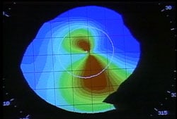
Corneal topography produces a detailed, visual description of the shape and power of the cornea. This type of analysis provides your doctor with very fine details regarding the condition of the corneal surface. These details are used to diagnose, monitor, and treat various eye conditions. They are also used in fitting contact lenses and for planning surgery, including laser vision correction. For laser vision correction the corneal topography map is used in conjunction with other tests to determine exactly how much corneal tissue will be removed to correct vision and with what ablation pattern.

Computerized corneal topography can be beneficial in the evaluation of certain diseases and injuries of the cornea including:
- Corneal diseases
- Corneal abrasions
- Corneal deformities
- Irregular astigmatism following corneal transplants
- Postoperative cataract extraction with acquired astigmatism
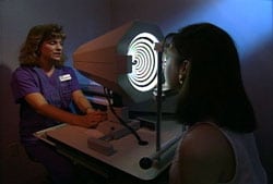
The corneal topography equipment consists of a computer linked to a lighted bowl that contains a pattern of rings. During a diagnostic test, the patient sits in front of the bowl with his or her head pressed against a bar while a series of data points are generated. Computer software digitizes these data points to produce a printout of the corneal shape, using different colors to identify different elevations, much like a topographic map of the earth displays changes in the land surface. The non-contact testing is painless and brief.


